

What is a Tracheotomy Procedure?
A tracheotomy procedure involves making an artificial hole called a stoma in the throat to restore a person’s ability to breathe when the airway is obstructed. Also referred to as a tracheostomy, how it is performed is determined by circumstances, dependent upon whether it is an emergency or a planned procedure. The intubation of an artificial breathing tube is normally a short-term situation, but can be used over the long-term as conditions require. As with any invasive procedure, a tracheotomy has some risk for complications, including scarring, infection and excessive bleeding. In most cases, an emergency tracheotomy is performed because of significant injury to the head, neck or upper torso. When a person’s ability to breathe is impaired, a secondary hole must be made to allow for respiration and air flow.
When it is performed in a hospital setting, a tracheotomy procedure involves using a general anesthetic. An incision is made just below the Adam’s apple near the thyroid, and penetrates the trachea, or windpipe. After making a small hole, a tracheostomy tube, or trach tube, is positioned into the hole. Sutures may be used to tighten the tissue around the tube and to prevent foreign material from getting into the hole. To keep the trach tube from being displaced or shifting, a small guard or plate is positioned around the exposed end of the tube and secured with an elastic strap or nylon. A planned tracheotomy is generally performed when a medical condition adds to the airway obstruction. Chronic conditions, such as paralysis and throat cancer, can make it necessary to have a tracheotomy. The most common contributory factors for airway obstruction are tracheal narrowing and impaired muscle function within the throat. Following neck surgery, it is not uncommon for a tracheotomy to be used to aid recovery.
What is a Tracheostomy Tube?
A tracheostomy tube is inserted into the stoma, or hole, and directly into the trachea to help keep the airway open for breathing. A tracheostomy tube typically consists of an inner tube, an inner cannula, an outer tube, an outer cannula, and an obturator. The obturator is used as a guide for the outer cannula during insertion. It is then removed after insertion, leaving the outer cannula in place to be a passageway for air to get through. In some cases, a single cannula tube is used with no inner tube, often for small children who require this procedure.
Certain tube varieties can be used depending on the severity and nature of the problem affecting the individual. A cuffed tracheostomy tube is often required to allow for mechanical ventilation in cases with severe respiratory failure. Generally, a tube cuff can be inflated different ways, it blows up like a small balloon, and allows the mechanical ventilation to work. Fenestrated tracheostomy tubes allow a person to regain the power of speech due to an opening on the tube that brings air in through an upper airway.
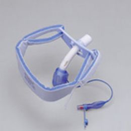
A tracheostomy tube holder is a device that can be used to stabilize a tracheostomy tube by securing it with hooks that are attached to the neck flange of the tube. The holders adjust around the neck and are normally made of a soft material to provide comfort and to prevent damage to the skin. A tracheostomy tube requires maintenance that directly relates to the severity of the problem. In some cases, a person will improve to the point where the tube is no longer needed, so it is removed, leaving a scar at the area of the stoma. But, most cases require tubes to be in place long-term. The initial tube is usually replaced at about two weeks after surgery, and if a tube is necessary for a long period of time, the individual may need to learn how to maintain the tube themselves. This could include suctioning out the trachea or even cleaning and changing the tube.
How do I Care for a Tracheostomy at Home?
Immediately after a tracheostomy, a person will communicate with others through writing until the healthcare provider gives instructions for other communication techniques. Also, do not remove the outer cannula unless the healthcare provider advises this. Use tracheostomy covers to protect the airway from outside elements, such as dust and cold air. Contact a healthcare provider or physician immediately if you’re experiencing an irregular heart rate, are feeling increased pain or discomfort, or if you’re having difficulty breathing and it is not relieved by the usual method of clearing the secretions. Also, contact a physician if secretions become thick, if crusting occurs, or mucus plugs are present.
Before leaving the hospital, a nurse should provide instructions on how to care for a tracheostomy tube. It should be done at least once a day after leaving the hospital. First, gather the needed supplies, wash your hands thoroughly, and sit or stand in a comfortable position in front of a mirror. Then, put on gloves, suction the trach tube, remove the inner cannula, hold it over the sink and pour antiseptic over and into it, cleaning it with a small brush. Thoroughly rinse it with normal water, then dry the inside and outside of the inner cannula completely, and reinsert and lock it in place. Remove the soiled gauze dressing around the neck and throw it away, and inspect the skin for redness, tenderness or drainage. If any of these occur, call your doctor. Use soaked swabs to clean the exposed parts of the outer cannula and the skin around the stoma. Dry the cannula and skin, change the trach tube ties, and then place a fine mesh gauze under the tracheostomy tie and neck-plate by folding or cutting a slit in it. Finally, remove the gloves and throw them away, and wash your hands with soap and water.
Hulet Smith, OT
Rehabmart Co-Founder & CEO
lb